This is default featured post 1 title

Go to Blogger edit html and find these sentences.Now replace these sentences with your own descriptions.This theme is Bloggerized by Lasantha Bandara - Premiumbloggertemplates.com.

This is default featured post 2 title

Go to Blogger edit html and find these sentences.Now replace these sentences with your own descriptions.This theme is Bloggerized by Lasantha Bandara - Premiumbloggertemplates.com.

This is default featured post 3 title

Go to Blogger edit html and find these sentences.Now replace these sentences with your own descriptions.This theme is Bloggerized by Lasantha Bandara - Premiumbloggertemplates.com.

This is default featured post 4 title

Go to Blogger edit html and find these sentences.Now replace these sentences with your own descriptions.This theme is Bloggerized by Lasantha Bandara - Premiumbloggertemplates.com.

This is default featured post 5 title

Go to Blogger edit html and find these sentences.Now replace these sentences with your own descriptions.This theme is Bloggerized by Lasantha Bandara - Premiumbloggertemplates.com.

Showing posts with label biology. Show all posts

Sunday, August 21, 2011

New Cure for Leukemia?


Leukemia is the cancer of white blood cells.
The Penn scientists targeted chroniclymphocytic leukemia (CLL) by hacking a harmless version of the HIV virus to hack T cells in order to kill cancer cells. In previous studies, the cancer-killing cells died out quickly after infusion, but in this study, the genetically engineered cells multiplied a thousand-fold and were sustained for over 4 months. 

Let's go over the study first.  Three patients with chemotherapy resistant tumors had their blood drawn, separated, modified, and cultured. These patients underwent lymphodepleting chemotherapy, and their blood was reinjected. Endpoint assays were conducted a month after reinjection.

They did the same thing in mice, and the cells of interest were sustained for over six months, although I'm not sure whether the same monthly cycle was repeated. It doesn't say. But the cells of interest reached levels of up to 95% of white blood cells, up from 2.3-4.46% (figure 2). After an initial decay with first-order kinetics, the CART19 cell numbers stabilized between three to six months after reinjection. The fact that the cell levels were sustained after four months is at least some evidence the body can remanufacture the CART19 cells on their own.

What is most remarkable, however, is that the cells of interest seem to be able to remanufacture themselves within the body. In the third patient, flow cytometry showed that there were CAR19-expressing T cells with an absence of B cells 169 days after infusion. This is remarkable, since, "previous studies have not demonstrated robust expansion, prolonged persistence, or functional expression of CARs on T cells after infusion."


Figure 2 from the study showing levels of CART18 cells after infusion. Click to enlarge.

"There were no significant toxicities observed during the 4 days after the infusion in any patient other than transient febrile reactions. However, all patients subsequently developed significant clinical and laboratory toxicities between days 7 and 21 after the first infusion...With the exception of B cell aplasia, these toxicities were short-term and reversible. Of the three patients treated to date, there are two complete responses and one partial response lasting greater than 8 months after CART19 infusion according to standard criteria." The only side effect these three patients suffered was fever. One was hospitalized for a week, and another went into remission for 10 months.

In fact, "one of the preclinical rationales for developing CAR+ T cells with 4-1BB signaling domains was a projected reduced propensity to trigger IL-2 and tumor necrosis factor–α (TNF-α) secretion compared to CAR+ T cells with CD28 signaling domains (7); indeed, elevated amounts of soluble IL-2 and TNF-α were not detected in the serum of the patients." The cells infused into the patients were designed specifically to avoid a cytokine storm and to circumvent the donor's immune system.

"In our preclinical studies, we found that large tumors could be ablated and that the infusion of 2.2 × 10⁷ CAR T cells could eradicate tumors composed of 1 × 10^9 cells, for an in vivo effector-to-target (E/T) ratio of 1:42 in humanized mice (8), although these calculations did not take into account the expansion of T cells after injection." In mice studies, billion cell tumors were ablated.

The three human patients had trillion cell tumors weighing around 1 kg before the infusion of CART19 cells. They all showed great progress, with the third patient surpassing others by 40:1. "Using the estimate of initial total tumor burden (1.3 × 10¹² CLL cells) and the observation that no CLL cells were detectable after treatment, we achieved a marked 1:93,000 E/T ratio. By similar calculations, an effective E/T ratio in vivo of 1:2200 and 1:1000 was calculated for UPN 01 and 02 (table S6). Therefore, a contribution of serial killing by CART19 cells combined with in vivo CART19 expansion of >1000-fold likely contributed to the powerful antileukemic effects mediated by CART19 cells."

The trials for the three patients were financed by Alliance for Cancer Gene Therapy.

(the study) and MSNBC.

Friday, August 19, 2011

Enumerating Bacteria In Lab


Serial dilutions allow us to do viable cell counts or total cell counts.

Serial dilution and plating can determine the amount of viable cells in a culture. Serial dilutions allow a discrete number of colonies of bacteria to grow, whereas concentrated cultures may contain billions of bacteria per milliliter. In serial dilutions, smaller dilutions are repeated in succession, and the dilutions can be multiplied to obtain the total dilution. Thus, serial dilutions are more practical than doing the total dilution in a single time. For example, if I have a 100 ml bacterial culture, I can add 1 ml of it to 99 ml of water, add 1 ml of the first dilution to 99 ml of water, and then add 1 ml of the second dilution to another 99 ml of water. I end up with a 10-2 X 10-2 X 10-2 = 10-6 dilution of the original bacteria culture. I can then plate .1 ml of the final dilution on growth medium. The goal is to dilute the culture so that, when plated, the number of bacterial colonies is discrete and each colony arises from one viable bacterial cell. We can use the number of viable cells in the undiluted culture by dividing apparent colony-forming units with the product of milliliters used and the dilution factor. For example, if 150 colony-forming units were counted on the plate that was streaked with .1 ml of the 10-6 dilution, there is about 150 / (.1 ml X 10-6) = 1.5 x 10⁹ bacteria/ml in the original culture. This method is useful because I am using only a small portion of the original culture, and large volumes of solution are not required for many-fold dilutions.

The Petroff-Hausser Counting Chamber can also be used to validate cell counts. The cell suspension is vortexed and a drop is applied to the chamber with a Pasteur pipette. Etched squares on the surface of the chamber representing specific areas and volumes are then examined under high magnification. Count the number of bacterial cells per chamber cell and multiply to obtain the concentration of cells per milliliter.

The turbidimetric method indirectly determines the quantity of insoluble particles in a liquid by comparing light transmittance in reference to a standard solution. A spectrophotometer shines a specific wavelength of light at the sample. Insoluble particles suspended in the sample will absorb and the incidental light, decreasing the amount of light transmitted to the photocell. Optical density is the measure of the turbidity of a solution, and it increases as the concentration and size of the particles increase. For example, as the concentration of bacteria reaches about 107 cells per ml, the liquid medium will appear cloudy or turbid.

Aλ= log10(Io/I) = εbc

The absorption of light is described by the Beer-Lambert Law, where A is absorbance, Io is the intensity of light incident on the sample, and I is the intensity of light transmitted through the sample. Beer’s Law states the optical density is proportional to the concentration of the compound in the solution, c, and the light’s path length, b. Thus, the concentration of bacteria in a pure culture can be determined if the molar absorbtivity, ε, and the path length, c, are known. OD600 refers to the optical density of a sample when the incident light has a wavelength of 600 nanometers. 

Sunday, August 14, 2011

Regulation of Morphology of Corn Smut, Ustilago maydis

Basidiomycota, in contrast to other fungi such as Ascomycota, produce basidia that yield four sexual spores called basidiospores. U. maydis is part of this phylum. Its mating-type is determined by a tetrapolar system with two unrelated loci, a and b. There are two idiomorphs for the a locus, a1 and a2. Haploid U. maydis cells have either the 4.5kb a1 locus with genes mfa1, pra1, and rfa2, or the 8kb a2 locus with genes mfa2, pra2, lga2, and rga2. mfa1 and mfa2 encode pheromone precursors, pra1 and pra2 genes encode pheromone receptors for the a2 and a1 pheromone, respectively. rfa2, lga2, and rga2 are thought to function within mitochondria. The pheromone encoded by one idiomorph will bind to the receptor of the opposite cell type, activating a signaling cascade that induces G2 arrest and the formation of conjugation hyphae. The b locus contains two genes, bE and bW, and regulates the switch to the pathogenic filamentous stage, as well as tumor induction and the formation of teliospores. The complex mating-type regulation is not specific to U. maydis, and other Basidiomycetes such as Schizophyllum commune and Coprinus cinereus.

Promycelium undergoes meiosis to produce saprophytic haploid cells. When in contact with corn, these sporidia exchange pheromones and become conjugative hyphae. These fuse to form a dikaryote, which is able to being intracellular invasion. Tumors are induced in which the fungi proliferates. Spores are formed and spread in the air and form a promycelium.

Higher fungi, like U. maydis, make ideal genetic models because they are easy to mate, transform, and select for. Observing metabolism, virulence, genotype is easier because they tend to be linked to readily apparent morphology. U. maydis’ relatedness to animal cells makes their study even more relevant to humans. Not only do does it have microtubule organization, nuclear migration, and nuclear envelop breakdown like in humans, U. maydis has homologues of Homo sapiens proteins that other, “higher” genetic models lack, such as Brh2, a BRCA2 (Breast Cancer Type 2 susceptibility protein) homologue. In vivo studies of Brh2 made it possible for geneticists to understand the function of BRCA2 in DNA repair and tumor suppression in humans. There is no doubt of U. maydis’ importance as a genetic model to study other complex mammalian cell processes. 

No pictures. No sources. Only excellence.

Friday, August 12, 2011

Question of the Day: Anti-HIV Antibody Edition


CN3D 4.3. Click to enlarge.


Jmol. Click to enlarge.
The first is modeled by the researchers, using the CN3 program. There's more colors available and the graphics look better. The one on the bottom is the same anti-HIV antibody in the Jmol program that I meddled with a little bit (ID: 3RPI).

This antibody mimics CD4 binding, locking onto to the spikes of HIV-1 so HIV-1 can't bind to (CD4) white blood cells. By characterizing its unique structure, researchers can design many novel antibodies that could efficiently inhibit the virus' entry into host white blood cells. 

 Random ethical hypothetical question here: You are granted the ability to cure HIV and AIDS and save millions of lives, but in order to use this ability, you must kill an innocent girl. You must choose between curing HIV and killing someone. What would you do? Explain your reasoning. I'll post my answer tomorrow.

Wednesday, August 10, 2011

Mesothelioma and Asbestos

First, the mesothelium. It's a frictionless monolayer lining that covers the internal organs. The luminal side has lots of microvilli that told fluids and proteins to allow intracoeolmic movement. It also helps leukocytes and other cells of the immune system to travel about in the fluid.


Mesothelium cells, with connective tissues.

How exactly does asbestos cause cancer? Well, it's made up of little tiny fibers that people inhale it. These fibers travel into the lungs and stick to its linings, damaging the membrane.  Intra-pleural inoculation of asbestos in rats have demonstrated that asbestos cause lesions within the lining, recruiting phagocytes and macrophages to the site of the lesion. The macrophages are part of the immune response, and they eat up cells which have asbestos in them. This in turn damages the macrophages and cause oxidative stress. Additionally, it is thought that smaller asbestos fibers can sometimes become entangled within the chromatin itself in the cell, and disrupt with the process of cellular division by interfering the packing and segregation of chromosomes. After many cycles of cellular division, the DNA damage accumulates. The subsequent damage induces the cell to undergo DNA repair, which is often error-prone. This is how asbestos damages the tissues, ultimately causing lesions to develop into a malignant tumor in the mesothelium.


Asbestos may interfere with cell division.
So why was asbestos use so widespread if the link between lung cancer and it was made in the early 19th century? Asbestos is a mineral that is heat-, friction-, and acid-resistant, easily obtained by mining, and easy to modify. These industrial merits are why companies today still incorporate asbestos into their products. Countries all over the world limit the use of asbestos. But whatever countries do to limit the use of asbestos, mesothelioma will still be a problem for years to come, because of there is long latency between asbestos exposure and the development of the disease. For example, the Japanese government expects the peak year for malignant mesothelioma to be in 2025. 

The widespread use of asbestos, the long latency period, the exclusive linkage between asbestos and malignant mesothelioma, and the fact that not all companies have enacted proper safety measures have opened the doors for a whole lot of lawsuits. People are seeking compensation, and in 1999, already 2 billion have been award to people. That's 2,000 million. No wonder there are lawyers and attorneys who specialize in mesothelioma cases.  Why not? Some mesothelioma lawyers have gotten recoveries of 3 million per victim. It's lucrative since not only is the five year survival rate very low (9%, so what's the point of a structured settlement?) and the treatment expensive, so many people were exposed to asbestos because of the American economy's emphasis on manufacturing (where contact with asbestos is most likely) after World War 2. On top of that, many companies that used asbestos were reluctant to get rid of asbestos, even when they knew the occupational hazards.

If you think you may have mesothelioma because you have the symptoms (weight loss, fever, cough, swelling due to fluid buildup) go get an MRI or a thoracoscopy (where they make an incision and put a camera to look into your chest).


Pleural mesothelioma tumor grows and effectively "shrinks" lung capacity. It affects 70% of patients.
Mesothelioma affects people from all walks of life. Steve McQueen died of a heart attack in Juarez, Mexico, after undergoing mesothelioma treatment. He was exposed to asbestos during his time in the United States Navy and during his long career as a race car driver. At the time, asbestos was used to insulate the piping on the boats and was also incorporated into  his racing suits.

Thursday, August 4, 2011

Perianal Sweat... Why You Should Use Deodorant

She looks hot! Very warm, indeed!
Eccrine sweat glands are found only in primates and reach their greatest development in humans. They are distributed all over the body, producing sweat for cooling. When we are warm, the hypothalamus tells the eccrine glands to start cooling us off as the sweat evaporates.

Apocrine sweat glands are larger and are limited to axilla (armpits) and perianal areas in humans. Because of the substances that are contained within apocrine sweat (e.g. protein, ammonia, lipids, chromogranins) it has a more thick and milky consistency than eccrine sweat. The healthy bacteria covering your body loves to munch away on apocrine sweat, and they multiply and divide when there is apocrine sweat. As the grow, they produce a lot of waste, which is what causes body odor.

Do you wear deodorant, or antiperspirant? There are some whisper going around about cancer and aluminum in many antiperspirants. The next post may be about a connection between the two!

Wednesday, August 3, 2011

TTAG In-Depth Article on Extremophilic Bacteria

Extremophilic bacteria thrive in extreme conditions. Bacteria in deep sea vents have to withstand high heat and/or high pressure conditions. They must have adapted to their extreme environment by having specific genes that encode more resilient proteins or have special metabolic pathways. What many researchers have done is analyze the genome of extremophilic bacteria, then compare their unique proteins with proteins from microbes that live in more ordinary environments.


A deep sea vent, known as a black smoker, where diverse extremophilic microbes thrive.
Bacteria from the deep sea were compared with shallow water counterparts by Dr. Lauro, such as Enterococcus faecalis with Carnobacterium. Dr. Reytor compared protein synthesis between extremophilic bacteria and temperate mesophiles such as common pathogens. Protein synthesis at these extreme conditions is made possible by deep sea extremophiles and other bacteria in extreme environments survive because their genome has additional genetic clusters, though their function is unknown. Dr. Costa, meanwhile, spent most of his time overviewing the metabolic pathways of these extremophiles, indirectly by measuring the concentrations of nitrite, nitrate, ammonia, and sulfide, as well as other organic and inorganic compounds within the native waters of the bacterium species. Costa's emphasis on the geochemistry and the setting of the bacteria will show where some thermophilic and extremophilic bacteria grow best. 

Carnobacterium is found deep in the Aleutian Trench, yet is genetically similar to Enterococcus faecalis, suggesting that minimal evolution was needed to make the switch. For example, P. profundum strain SS9 and strain 3TCK have different bathytypes but show great synteny when their genome was sequenced (Lauro 20). 


E. faecalis (pictured above) and Carnobacterium are genetically similar!
“An essential cellular process inhibited by hydrostatic pressure is protein synthesis. Hydrostatic pressure induces the synthesis of a number of ribosomal and heat-shock proteins in Lactobacillus and Escherichia Coli.” However, many bacteria found in the vents are the exact opposite, having “an SOS response, with heat-shock genes being over-expressed at atmospheric pressure due to partial loss-of-function of the ribosomal units to create folded proteins” (Lauro 19). The most major difference genetically speaking between deep sea bacteria and shallow water ones were genes that coded for flagella synthesis and motility, as these are the most affected by pressure. One difference that was found was that deep sea vent bacteria had greater intergenic spacers. Additionally, a greater percent of rRNA copies were found on deep bathytypes than shallow water bacteria. Many genes were found in the genome of deep bathytypes coded for cell membrane unsaturation, as well as photolyase (Lauro 20). Photolyase repairs the cyclobutane prymidine dimers caused when blue light strikes DNA. When bacteria are this deep in the water, this gene is not necessarily needed to be expressed. 


Lipases from Caldanaerobacter subterraneus subspecies tengcongenesis, and Thermoanaerobacter thermohydrosulfuricus were cloned and expressed in Escherichia coli. Lipase hydrolysizes and synthesizes long-chain acylglycerols, with a wide range of use in dairy, pharmaceutical, and the food industry in general. For instance, “the lipase from Thermoanaerobacter thermohydrosulfuricus were extremely S-stereoselective towards esters of secondary alcohols (Royter 769).” This is useful because it will create extremely pure compounds only. These lipases are extremely resistant to high temperature and were active across a wide range of pH. This is why lipase from extremophilic bacteria are so useful, because they can survive above 70 and 80 degrees. These enzymes are called “extremozymes.” These extremozymes are better studied when recombined with mesophilic bacteria, so they have been cloned and expressed in Escherichia coli, instead of their extremophilic microbe cousins. 


Thermoanaerobacter thermohydrosulfuricus was isolated from Solar Lake, while Caldanaerobacter subterraneus subspecies tengcongenesis was taken from a Chinese hot spring. To make them reproduce, the agar they chose had to have special nutrients, specifically metal halides (Royter 771). They were incubated at 65 degrees, almost twice the optimal growth temperature of a typical mesophilic bacteria. Escherichia coli was cultivated in Luria-Bertani agar, with tryptone and 1% sodium chloride. Escherichia coli was transformed using heat-shock, and the DNA fragments that were cut up by HindIII and BamHI were analyzed with the PCR technique (Royter 771). 

To determine what the lipase used as active sites, various chemical compounds were added into the culture. If the compounds inhibited the enzyme, that means the compounds either directly or indirectly competed with the normal substrate. Therefore, the active site must contain some of these groups. A spectroscope was used to see the effects of metal halides and other chemical compounds on purified lipase. Lipase was added with these compounds, then after a short amount of time, residual activity of the enzyme was measured (Royter 772). (Royter 777). “Serine and thiol groups are part of the active site for these enzymes (Royter 769).” As a result of this research, it was concluded that due to their active sites, the lipase and other enzymes of this nature came from mesophilic and psychrotrophic bacteria. 

To determine the optimum pH for the operation of the lipase that was synthesized with cloned genes, turbidity was measured. Samples of lipase were faced to see how much copper soap could be used to create fatty unsaturated acids. Also, samples of the lipase with the lipase were subjected to acetic acid and sodium hydroxide for 70+ minutes (Royer 772). This questioned whether or not the lipases were stable with strong bases. 

Also, the enzyme was subjected to two cycles of thawing and freezing. Some reduction was reported, but it was fairly resistant, with 70-80% remaining (Royter 775). 

The enzyme was then placed within different solutions of esters to see the relative rate of hydrolysis. The enzymes were most effective with secondary alcohols (Royter 775). 

After the transformation of Escherichia coli, lipase activity was measured in a supernatant without cells, but with cell lyase and para-nitrophenyl palmitate. This cell-free supernatant was created by precipitating heat, hydrophobic interaction, and then gel-filtering the substance. Residual activity is what it is called freed from the cell. 

The results and conclusions were that over 17 amino acids were found to be similar in the terminal amino acid of the lipases between Thermoanaerobacter thermohydrosulfuricus and Caldanaerobacter subterraneus subspecies tengcongenesis. The genes were then sent through PCR and amplified. The optimum temperature for the lipase was 75 degrees, with a pH of about 8.0 (Royter 775). They remain active up to 90 degrees, as well. A 10% decrease in Thermoanaerobacter thermohydrosulfuricus lipase activity was measured when it was incubated at 85 degrees Celsius for 50 minutes. However, Caldanaerobacter subterraneus subspecies tengcongenesis saw an 80% reduction in residual lipase activity after the same incubation. These enzymes has an activity peak similar to the peaks with regular enzymes, with it skewing towards the right, with a sharp decline past the optimal temperature as the enzyme is denatured. 
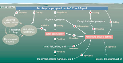

Microbial loops play an important role in marine ecosystems, digesting marine snow. (click to enlarge!)
Extremophiles are important because they represent more than half of all sea microbes live deep in the ocean depths (Lauro 15). They survive by reducing inorganic compounds, without the presence of oxygen, in the presence of great pressure. Microorganisms that are found in hot springs need to be able to thrive in harsh environments, as they are usually around 73 degrees (Costa 447). As said before, protein synthesis is often inhibited by high temperature or high pH, so the genetic differences of Thermoanaerobacter thermohydrosulfuricus and Caldanaerobacter subterraneus subspecies tengcongenesis and mesophilic Escherichia coli are notable when coding for exoproteins. The transformation was successful and Escherichia was able to produce these novel proteins, usually from bacteria that are completely anaerobic and use sulfur in their metabolic pathway in reduction-oxidation reactions. 

Antranikian G. (2008) Industrial relevance of thermophiles and their enzymes. In: Rob et al. (eds) Thermophiles—biology and technology at high temperature. CRC Press Boca Raton. Pp 113-160. 

Costa C. Kyle, Navarro B. Jason, Shock L. Everett (2009) Microbiology and geochemistry of great boiling and mud hot springs in the United States Great Basin. Extremophiles 13:447-459. 

Lauro Frederico, Bartlett Douglas. (2007) Prokaryotic lifestyles in deep sea habitats. Extremophiles 12:15-25 

Royter Marina, Schmidt M., C. Elend. (2009) Thermostable Lipases from the thermophilic anaerobic bacteria Thermoanaerobacter thermohydrosulfuricus SOL1 and Caldanaerobacter subterraneus subsp. Tengcongenesis. Extremophiles 13:769-783.

Saturday, July 30, 2011

The lac Operon Explained [Updated]

Click to enlarge Photo: NIH 
The lac operon is a favorite of microbiology and biology textbooks, used to explain transcription, regulation, and catabolism. Here's my quick synopsis of the regulation involved, making sure the cell only transcribes genes that encode lactose-eating enzymes only when it is physiologically necessary. But first, a GENETICS crash course in biology:

An operon is a group of genes transcribed as a single unit. It's like buying Orange Box. There's five separate games, but they're all purchased at once in a neat box. The same thing with an operon. If the operon is ON, you get all of the genes activated. If the operon is OFF, none of the genes are activated.

RNA polymerase is a protein that reads DNA molecules in the cell to make RNA. This RNA is then read again by ribosomes to direct the synthesis of specific proteins..

The lac operon is just a string of DNA with two regulatory sites on the DNA and three structural genes (but we'll only talk about two here). The first of the two regulatory sites is the promoter, where the RNA polymerase binds to read the operon to make an RNA copy of it. The second regulatory site is called the operator, where the repressor binds to the operon. The promoter and operator overlap, so if the operator site is occupied by the repressor, RNA polymerase can't access the promoter to start reading the rest of the operon. Imagine the repressor and RNA polymerase as two brothers, and think of the operator and promoter sites as a chair that just big enough for one of two brothers to sit in. One of the brothers, the repressor, is lazy and if he sits in the chair he's going to nap. If the other brother, RNA polymerase, gets to sit in the chair, he's going to be productive and a ton of protein's going to be made. These brothers aren't going to share the chair; either the repressor or RNA polymerase sits in it, but not both. If the repressor binds to operator, the operon is in the OFF position. No genes in the operon are being transcribed, no proteins are being made in that operon. If RNA polymerase binds to the promoter, the operon is in the ON position, and the structural genes are being transcribed. What decides who gets the chair, then?

Turns out the lazy repressor brother is allergic to lactose, so if there's lactose around, he dies and RNA polymerase gets to sit in the chair. In the presence of lactose, the repressor is inactivated, and as a result, the RNA polymerase is free to bind to the promoter of the operon and begin transcription of lacZ and lacY genes, which will be subsequently translated into galactosidase and permease proteins, respectively. These proteins allow the cell to break down lactose and use it as an energy source. First, permease must to allow lactose to enter the cell. Then galactosidase is required to break down lactose, a two ring sugar, into galactose and glucose, both single ringed sugars. Both proteins are required if the cell is to use lactose effectively as a source of energy. The fact that RNA polymerase only gets access to the promoter ("the chair") in the presence of lactose means the cell will only produce proteins required for lactose breakdown when there is actually lactose to break down. Otherwise, the repressor binds to the operator, and neither permease nor B-galactosidase is made.

In addition to negative regulation by the repressor, the lac operon is positively regulated by cAMP, cyclic adenosine monophosphate. E. coli doesn't really like lactose. Positive regulation by cAMP ensures the cell will use lactose as a carbon source only in the absence of glucose, which is the preferred sugar for most cells because it is more efficient metabolically. This is known as catabolite repression. Glucose and cAMP levels are inversely proportional; the higher the concentration of glucose, the lower the concentration of cAMP. Only under low concentrations of glucose and high concentration of cAMP will the lac operon be activated and galactosidase and permease be produced. 

So there you have it. The operon is on only in the presence of lactose, when lactose binds and inactivates the repressor. The operon is on (to a much higher degree) when the cell is "starving" because there's no glucose around, and so the cell uses lactose as a backup carbon source.

Tuesday, July 26, 2011

Origin of Life?

courtesy of dailygalaxy.com
There's a fascinating article in Nature (here's the article, courtesy of the University of Texas) on how life started on Earth, and the evidence in rRNA that suggests it came from deep hydrothermal vents in the ocean. The article here focuses on the similarities between the metabolism of autotrophs and the geochemical reactions that take place in the vents. The reactive gases, dissolved elements, the pH, and thermal gradients at the vents are suitable for the development of life. Given that the same type of environment was very common in early Earth's oceans, it is likely that the microbes that thrive at the vents could be closely related to the first microbes on Earth, despite oxic conditions and carbonate chimneys in modern vents. The vents in early Earth were anoxic, and were FeS-rich in comparison. Anaerobic methane-oxidizing microbes' biological pathways were compared to the geochemical process of serpentinization, where water and bicarbonate reduce Fe2+ in hydrothermially altered mantle rock to produce serpentine and hydrogen gas. Hydrogen gas is an electron source that can, in turn, reduce carbon dioxide to methane in a process similar to the biochemical pathways of local methane-oxidizing microbes. This comparison is important because if life did arise from systems like LCHF, serpentinization could have been an evolutionry precursor to the first biochemical pathways. To support the possibility of life originating at LCHF-like systems, the authors mentioned that the pH and thermal gradients at the LCHFs would allow high concentration of biological precursors to form, and that they could be used to explain the origins of chemiosmotic coupling in modern autotrophs. 


In contrast to the anoxic oceans of the Hadean era, today's oceans have plenty of oxygen. Thus the chemical of composition of the LCHFs and the vents would be different from the oceans of early Earth. The presence of oxygen means very different microbes would exist then, compared to now. How do aerobic microbes in the hydrothermal vents, figure in finding the origins of life? Are they of any special interest, or should further research focus on methanogens only? How did lipid bilayers, a sure prerequisite for life, come about? Most of the compounds discussed in the article are very simple and small.

Monday, July 25, 2011

Why Cancer Is Going To Be Difficult To Beat

As homemakesgames said in the last QOTD, cancer is very complicated. There are so many different types of cancers. There is not going to be a single cure for all cancers. Cancer is not a single mutation in our cells, it's a whole bunch of possible mutations that leave the cell unable to receive feedback from its environment or properly repair damaged DNA. Cancerous cells grow blood vessels to bring nutrients in from surrounding, healthy tissue, disrupting them. Not only do they quickly invade healthy tissue, they have figured out a way to cheat death. Ignoring all kinds of growth inhibitors from the rest of the body, cancerous cells circumvent limitations on how many times its DNA can be copied. They have figured out ways to deactivate programmed limits in its own lifespan. This process can occur in many ways, which makes finding a common cure near impossible. If there is only one thing all cancers have in common, it is that, without treatment, their host cannot sustain them.

Twitter Delicious Facebook Digg Stumbleupon Favorites More